8. Nanoparticle-based sensors

Sensors based on nanoparticles are particularly attractive for intra- and extracellular imaging. They the flexibility of water-soluble indicators with robustness of optical sensors, since the polymeric or sol-gel matrix provide protects the indicator from undesired interferences (e.g. from interaction with proteins and other biological substances which may significantly change sensing properties). Such shell also reduces the toxicity of the probe in case of intracellular measurements. Additionally, functional groups on the surface of the nanoparticles can facilitate cell penetration. Finally, small size of nanoparticles ensures virtually real-time response. Due to these properties, nanoparticle-based sensors are also attractive for measurements in samples of small volumes such as respiration vials or microplate wells. We presented a versatile concept for designing optical nanosensors based on commercially available poly(styrene-block-vinylpyrrolidone). [47] These neutral beads were swollen by addition of water-mixable organic solvent (e.g. tetrahydrofuran) and the solution of a lipophilic indicator was added. The slow removing of the solvent under vacuum resulted into incorporation of the indicator into the core of the nanoparticle (oxygen, temperature probes). Alternatively, using ethanol:water mixture as a solvent (in which the nanoparticles do not swell) resulted into incorporation of the indicator into the shell of the particle (pH, ion sensors). In the first work, we demonstrated that preparation of oxygen, pH, chloride and copper nanosensors was possible. In the further work [48] we a variety of oxygen nanosensors utilizing the above concept was prepared. The nanobeads incorporated different classes of the dye and therefore have high flexibility in choice of spectral properties (to ensure good compatibility with light sources and detectors) and sensitivities (e.g. by using palladium(II) porphyrin for monitoring low oxygen and platinum(II) porphyrins for measurements at physiological concentrations). We also incorporated a variety of state-of-the-art pH indicators into poly(stryrene-block-vinyl pyrrolidone) beads. [49] Fluoresceins bearing electron-donating and electron-withdrawing substituents were shown to cover broad dynamic range when incorporated into the nanobeads. Later, nanoprecipitation was explored as simple and versatile strategy for preparation of nanosensors for various analytes from different polymers (Fig. 8.1). [50] Briefly, a polymer and a dye are dissolved in an organic solvent, which is mixable with water. Then the solution is either rapidly or slowly mixed with water (slow addition of water into the solution of the polymer or vice versa). This results in formation of the nanoparticles; organic solvent is removed via evaporation or dialysis. We demonstrated that a variety of commercially available polymers can be used and those combining hydrophobic backbone (polystyrene, polymethylmethacrylate) with some charged groups (carboxyl-groups, quaternary ammonium groups) are particularly suitable for preparation of small (∅ 100 nm) stable nanoparticles. Nanosensors for oxygen and temperature were fabricated by immobilization of a lipophilic porphyrin indicator into Rl-100 beads and a lipophilic Eu(III) complex into gas blocking poly(vinylidene chloride-co-acrylonitrile) beads. On the other hand, more hydrophilic fluorescein indicators have been incorporated in hydrophilic hydrogel beads to produce pH nanosensors. As was demonstrated in the application work, the nanosensors based on polymethylmethacrylate bearing quaternary ammonium groups (Rl-100) are excellently suitable for intracellular monitoring of molecular oxygen in various cell types, [51] whereas those based on polymethylmethacrylate bearing negatively-charged carboxylic acid groups can selectively stain neurocells. [52] The nanosensors presented excellent analytical tools for monitoring of oxygen dynamics in various cell types.
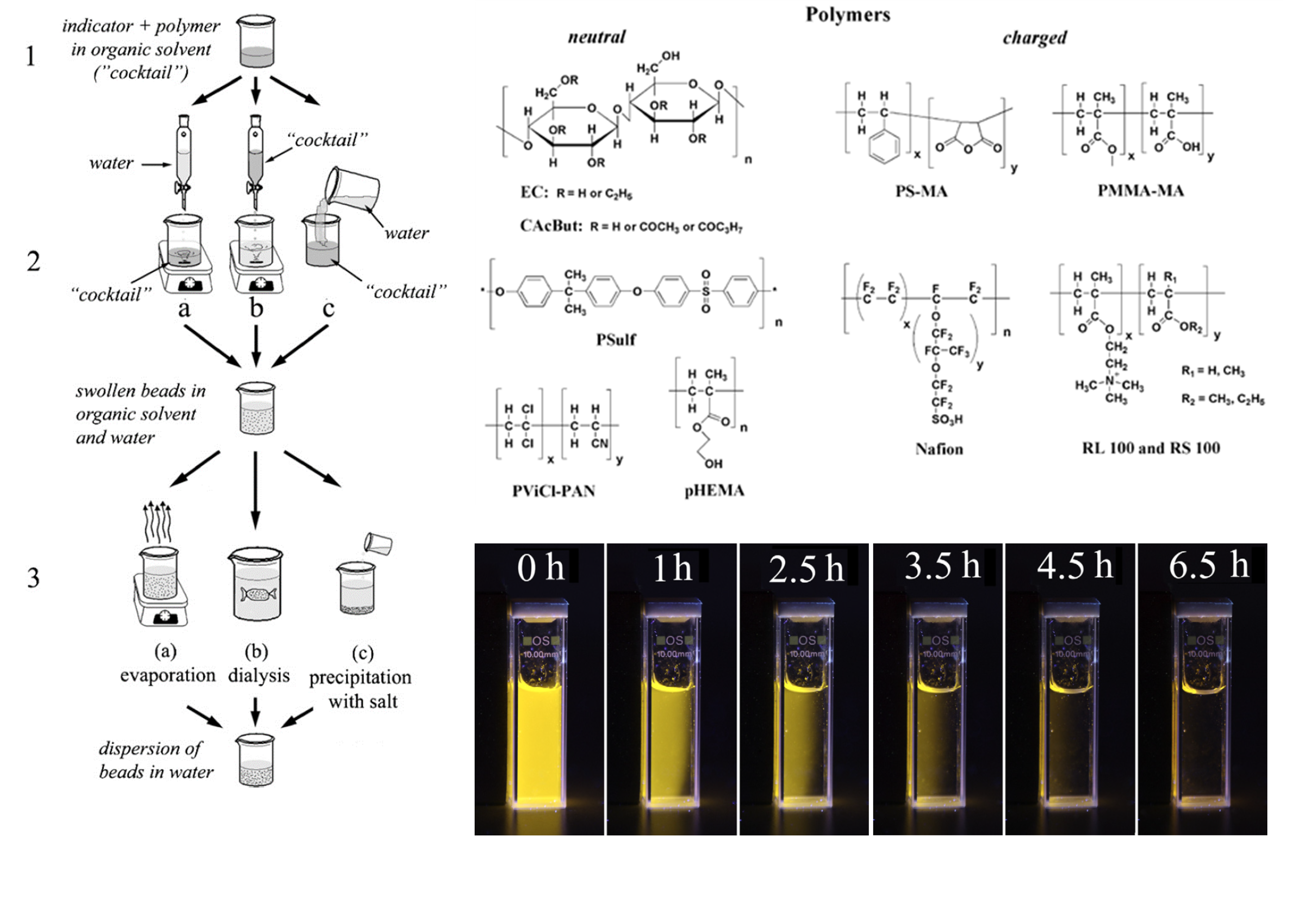
Figure 8.1. Left panel: illustration of the procedure for preparation of optical nanosensors via nanoprecipitation; upper right panel: chemical structures of the investigated polymers; lower right panel: photographic images of aqueous dispersion of magnetic oxygen sensitive nanoparticles under UV excitation during collection with a magnet. Reproduced from ref. [50].
Two-photon microscopy (2-P) enjoys increasing popularity among imaging techniques due to higher resolution, absence of background fluorescence and much better penetration of NIR light into tissues. Unfortunately, conventional dyes are poorly suitable for this technique due to very low 2-P absorption cross-sections. On the other hand, highly conjugated systems are known to have much better 2-P absorption cross-sections. Thus, we prepared an improved version beads for intracellular monitoring based on Rl-100 polymer. [53] Poly(9,9´)-alkyl fluorene was used as a 1-P and 2-P antenna (10 % wt. in respect to Rl-100) for oxygen indicator PtTFPP. Upon excitation with NIR light, bright oxygen-dependent luminescence of the Pt(II) porphyrin was observed. Residual blue fluorescence from the conjugated polymer enabled ratiometric referenced imaging. In the further work [54] we prepared series of conjugated polymers with covalently grafted oxygen indicators (Fig. 8.2). Polyfluorene or poly(fluorene-alt-benzothiadiazole) acted as a 1-P and 2-P antenna for red emitting Pt(II) di(pentafluorophenyl)diphenylporphyrin (Series I) and NIR-emitting Pt(II) tetraphenyltetrabenzoporphyrin (Series II), respectively. We also introduced positively-, negatively- or both positively and negatively-charged groups to enhance particle stability in aqueous media and to ensure cell-penetration of the beads. The beads were prepared via a nanoprecipitation method. A very efficient FRET from the conjugated polymer antenna to the oxygen indicator was observed in the nanoparticles (Fig. 8.3). Residual fluorescence from the conjugated polymer was used for reference purposes. Importantly, the beads showed bright luminescence also under 2-P excitation, making them particularly suitable for oxygen imaging in tissue spheroids. Overall, the brightness of the beads under 2-P excitation was significantly higher than that of the Rl-100 beads containing 10 % wt. of polyfluorene antenna. Later, we discovered that the zwitter-ionic beads readily adsorb on glass surfaces forming an oxygen-sensitive coating. Even relatively thin layers resulted in sufficient luminescence signal upon excitation in the conjugated polymer antenna which was used for post-modification of glass microfluidic chips. [55] Importantly, oxygen-sensitive coating was introduced in a very simple procedure (incubation with the dispersion of the beads and washing) which is advantageous to commonly used methods where chip bonding is required after integration of the sensing spot. We demonstrated potential applicability of the modified chips for measuring oxygen and for monitoring of activity of enzyme-loaded microparticles.
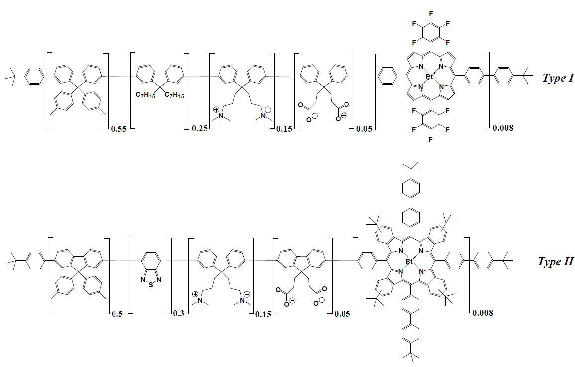
Figure 8.2. Chemical structures of the conjugated polymers used for preparation of oxygen nanosensors for 1-P and 2-P imaging. Reproduced from ref. [54].
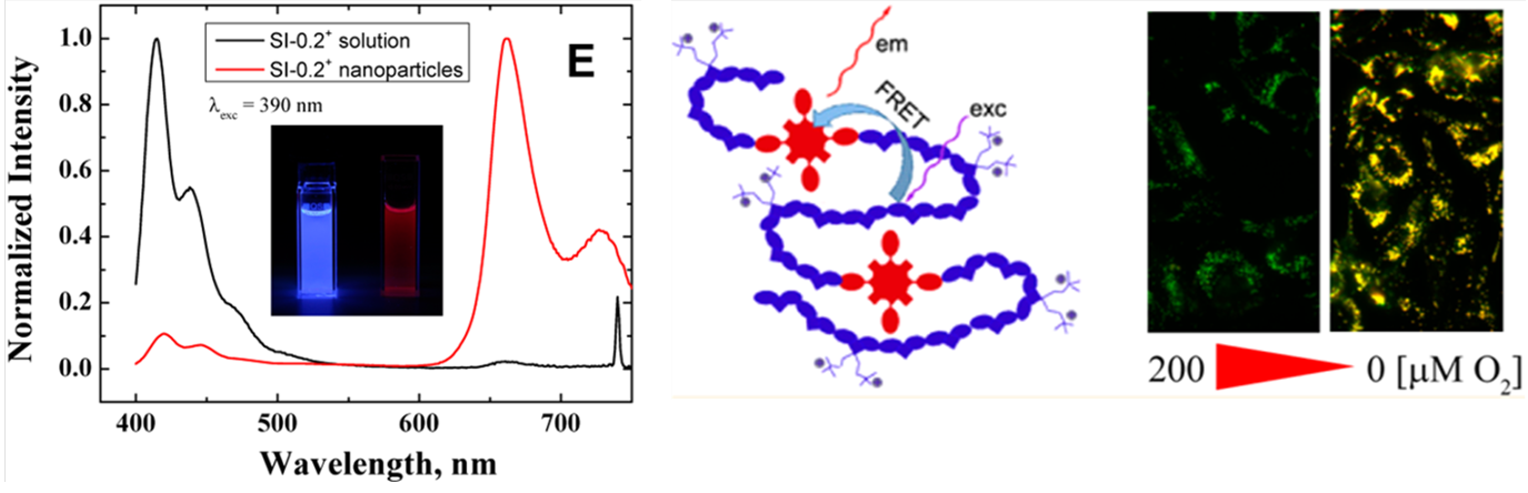
Figure 8.3. Left: emission spectra of the conjugated polymer dissolved in tetrahydrofuran and in the form of the nanoparticle dispersion in water illustrating efficient energy transfer from the conjugated polymer backbone to the oxygen indicator (middle); example of ratiometric intracellular measurement. 

References:

[43] Borisov, S. M.; Neurauter, G.; Schroeder, C.; Klimant, I.; Wolfbeis, O. S. Modified Dual Lifetime Referencing Method for Simultaneous Optical Determination and Sensing of Two Analytes. Applied Spectroscopy 200660, 1167–1173. [44] Schröder, C. R.; Polerecky, L.; Klimant, I. Time-Resolved PH/PO2 Mapping with Luminescent Hybrid Sensors. Analytical Chemistry 200779 (1), 60–70. [45] Vasylevska, G. S.; Borisov, S. M.; Krause, C.; Wolfbeis, O. S. Indicator-Loaded Permeation-Selective Microbeads for Use in Fiber Optic Simultaneous Sensing of PH and Dissolved OxygenChemistry of Materials 200618 (19), 4609–4616. [46] Kocincova, A. S.; Borisov, S. M.; Krause, C.; Wolfbeis, O. S. Fiber-Optic Microsensors for Simultaneous Sensing of Oxygen and PH, and of Oxygen and Temperature. Analytical Chemistry 200779 (22), 8486–8493. [47] Borisov, S. M.; Krause, C.; Arain, S.; Wolfbeis, O. S. Composite Material for Simultaneous and Contactless Luminescent Sensing and Imaging of Oxygen and Carbon DioxideAdvanced Materials 200618 (12), 1511–1516. [48] Borisov, S. M.; Seifner, R.; Klimant, I. A Novel Planar Optical Sensor for Simultaneous Monitoring of Oxygen, Carbon Dioxide, PH and TemperatureAnal Bioanal Chem 2011400 (8), 2463–2474. [49] Borisov, S. M.; Mayr, T.; Klimant, I. Poly(Styrene-Block-Vinylpyrrolidone) Beads as a Versatile Material for Simple Fabrication of Optical Nanosensors. Analytical Chemistry 200880 (3), 573–582. [50] Borisov, S. M.; Klimant, I. Luminescent Nanobeads for Optical Sensing and Imaging of Dissolved Oxygen. Microchim Acta 2008164 (1–2), 7–15. [51] Borisov, S. M.; Herrod, D. L.; Klimant, I. Fluorescent Poly(Styrene-Block-Vinylpyrrolidone) Nanobeads for Optical Sensing of PHSensors and Actuators B: Chemical 2009139 (1), 52–58. [52] Borisov, S. M.; Mayr, T.; Mistlberger, G.; Waich, K.; Koren, K.; Chojnacki, P.; Klimant, I. Precipitation as a Simple and Versatile Method for Preparation of Optical Nanochemosensors. Talanta 200979 (5), 1322–1330. [53] Fercher, A.; Borisov, S. M.; Zhdanov, A. V.; Klimant, I.; Papkovsky, D. B. Intracellular O2 Sensing Probe Based on Cell-Penetrating Phosphorescent Nanoparticles. ACS Nano 20115 (7), 5499–5508. [54] Dmitriev, R. I.; Borisov, S. M.; Kondrashina, A. V.; Pakan, J. M. P.; Anilkumar, U.; Prehn, J. H. M.; Zhdanov, A. V.; McDermott, K. W.; Klimant, I.; Papkovsky, D. B. Imaging Oxygen in Neural Cell and Tissue Models by Means of Anionic Cell-Permeable Phosphorescent Nanoparticles. Cell. Mol. Life Sci. 2014, 1–15. [55] Kondrashina, A. V.; Dmitriev, R. I.; Borisov, S. M.; Klimant, I.; O’Brien, I.; Nolan, Y. M.; Zhdanov, A. V.; Papkovsky, D. B. A Phosphorescent Nanoparticle-Based Probe for Sensing and Imaging of (Intra)Cellular Oxygen in Multiple Detection ModalitiesAdvanced Functional Materials 201222 (23), 4931–4939. [56] Dmitriev, R. I.; Borisov, S. M.; Düssmann, H.; Sun, S.; Müller, B. J.; Prehn, J.; Baklaushev, V. P.; Klimant, I.; Papkovsky, D. B. Versatile Conjugated Polymer Nanoparticles for High-Resolution O2 Imaging in Cells and 3D Tissue ModelsACS Nano 20159 (5), 5275–5288. [57] Lasave, L. C.; Borisov, S. M.; Ehgartner, J.; Mayr, T. Quick and Simple Integration of Optical Oxygen Sensors into Glass-Based Microfluidic Devices. RSC Adv. 20155 (87), 70808–70816. [58] Sun, S.; Ungerböck, B.; Mayr, T. Imaging of Oxygen in Microreactors and Microfluidic Systems. Methods Appl. Fluoresc. 20153 (3), 034002.