2. Glucose sensors

Optical glucose sensors of several types are known. Most common systems explore the sugar-binding properties of concanavalin A or boronic acids attached to a chromophore or make use of oxidation of glucose catalysed by glucose oxidase (Fig. 2.1). The enzymatic sensors are distinguished by excellent selectivity. The enzymatic reaction can be detected via heat production, decrease of pH, production of hydrogen peroxide or consumption of oxygen. The latter offer a very robust read-out and have been transducers of choice in the glucose sensors developed in our group. Miniaturised fiber-optic glucose sensors prepared in our lab (Fig. 2.2) enabled continuous measurement of glucose in subcutaneous tissue. [17,18] The sensor array consists of two oxygen optodes wherein one optode contained immobilized glucose oxidase and a diffusion barrier for adjustment of the dynamic range of the glucose sensor for physiologically relevant conditions. The glucose signal is a difference between pO2 of both optodes and allows for compensation of oxygen fluctuations in intercellular fluid. It was demonstrated that the optimized sensor was free of oxygen fluctuations in the expected pO2 range between 53 and 95 hPa and the sensor output was linear in the physiologically relevant glucose range up to 20 mM.  
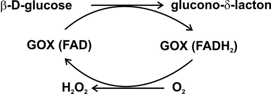
Figure 2.1. Mechanism of enzymatic oxidation of glucose. Reproduced from ref. [17,18].
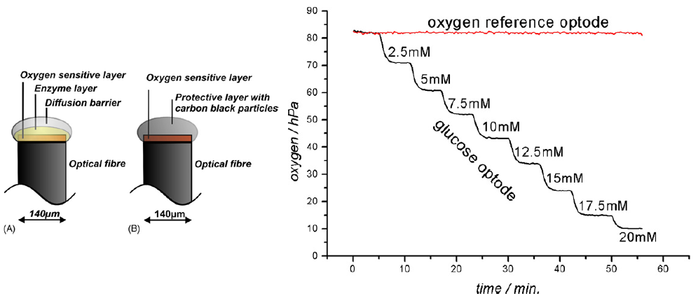
Figure 2.2. Glucose microoptodes for continuous glucose monitoring in subcutaneous tissue: left – schematic representation of the optode structure, right – example of the calibration curve. Reproduced from ref. [17,18].
Preparation of bright NIR oxygen indicators enabled design of a subcutaenous optical glucose sensor (Fig. 2.3). [19] The glucose sensor was coated onto an outer wall of a catheter and the read-out was performed through the skin. Additional oxygen sensor was added to enable compensation of the glucose sensor for oxygen fluctuations. Importantly, both oxygen indicators were carefully selected in order to (i) be excitable with the red light and have NIR emission to minimize the light lost in the tissue due to scattering and absorption and (ii) enable separate excitation of the dyes and collection of the emission to minimize the optical cross-talks between the analytes. Particularly, Pt(II) complexes with tetraphenyltetrabenzoporphyrin and aza-tetrabenzoporphyrin were used in the oxygen and glucose sensors, respectively. A dedicated read-out device (Fig. 2.3) was designed to enable interrogation of both sensors in two different channels. In-vivo measurements in pigs demonstrated good correlation of reference blood glucose levels and glucose values obtained with the presented sensor system. 
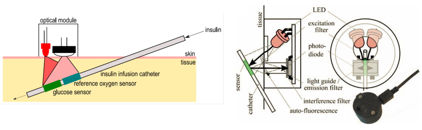
Figure 2.3. Schematic representation of the catheter-based subcutaneous glucose sensor (left) and the components of the read-out device designed for interrogation of glucose and oxygen sensors (right). Reprodcued from [19].

References:

[17] Pasic, A.; Koehler, H.; Klimant, I.; Schaupp, L. Miniaturized Fiber-Optic Hybrid Sensor for Continuous Glucose Monitoring in Subcutaneous TissueSensors and Actuators B: Chemical 2007122 (1), 60–68. [18] Pasic, A.; Koehler, H.; Schaupp, L.; Pieber, T. R.; Klimant, I. Fiber-Optic Flow-through Sensor for Online Monitoring of Glucose. Anal Bioanal Chem 2006386 (5), 1293–1302. [19] Nacht, B.; Larndorfer, C.; Sax, S.; Borisov, S. M.; Hajnsek, M.; Sinner, F.; List-Kratochvil, E. J. W.; Klimant, I. Integrated Catheter System for Continuous Glucose Measurement and Simultaneous Insulin Infusion. Biosensors and Bioelectronics 201564, 102–110.