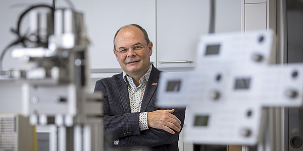
The aorta, the main artery, is responsible for transporting our blood from the heart to the furthest corners of our body. And it can become seriously ill. Gerhard A. Holzapfel, head of the Institute of Biomechanics at Graz University of Technology (TU Graz), has focused much of his research on atherosclerosis. Six years ago he found a new focus: aortic dissection – a disease that affects around six in 100,000 people and is still little researched due to its relative rarity.
A healthy aorta consists of several individual wall layers that are connected to each other. In an aortic dissection, the wall layers in the aorta become detached. This creates a kind of false lumen, in other words – a cavity or a second pathway parallel to the original true lumen where blood flows. The aorta can narrow or tear, or thrombi can form and detach. Funded by TU Graz, researchers from the fields of biomedical engineering, mathematics, fluid mechanics, electronics, computer science, civil engineering and mechanical engineering have been investigating this disease and possible diagnostic options over the past six years in the lead project “Mechanics, modelling and simulation of aortic dissection”. “The aorta is incredibly interesting from an engineering point of view,” says Gerhard A. Holzapfel. “It has a wall that consists of an elastic solid whose microstructure we can analyse using biophysical and microscopic methods. The blood inside the wall follows the laws of fluid mechanics and is also an excellent conductor of electricity – so I can observe how a very weak electric field affects conductivity and blood flow.”
From the basics to simulation
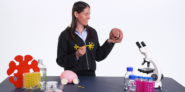
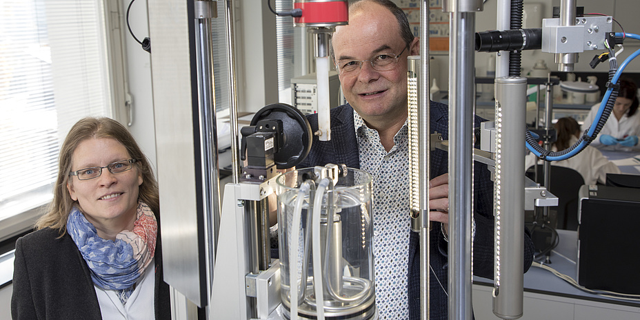
Selda Sherifova deals with the basics of the aortic wall. The post-doc is analysing wall samples of diseased aortas provided by the Medical University of Graz. She cuts out small areas from the samples, which she examines in the laboratory using various tests to determine their elasticity and behaviour under the influence of force. “If the aorta is diseased, certain proteins change. This can cause the aorta to lose its structure and eventually break. It was incredible to see this for the first time. Normally, the individual layers are very well connected – but if the aorta is diseased, the layers can detach even at the slightest contact,” explains the researcher. As part of her research in recent years, she has generated data on the mechanical behaviour of the aortic wall under various stress conditions, which is used for simulations and modelling to better predict both the course of the disease and the effects of various medications.
Malte Rolf-Pissarczyk is one of the people responsible for these simulations and models. He uses the previously generated data and CT images of patients to create patient-specific geometries of the aorta and subsequently model them using numerical methods. “This gives us an impression of how the blood flow in the aorta works. On the basis of this, we can then look at parameters such as wall tension, wall deformation and blood flow throughout the cardiac cycle. These parameters change when the aorta remodels itself in the event of illness, for example.”
Start-up for new diagnostic tool
The research results from the lead project are now to be made available to the general public in a promising spin-off from TU Graz. The arterioscope, a new type of diagnostic system, was developed to help medical staff recognise aortic diseases.
In impedance cardiography, an examination method similar to the electrocardiogram (ECG), weak electrical fields are applied to the body. Electric current takes the path of least resistance – in the case of the aorta, not the aortic wall, but the blood. At the same time, the current flow depends on the direction in which the blood flows. With every heartbeat, the aorta generates rhythmic pressure waves that change the conductivity. “If these factors change, this can indicate disease,” explains Sascha Ranftl, who works together with Vahid Badeli in the new start-up. The aim is to jointly create a database that can be used by conventional diagnostic methods to automatically identify pathological changes in the aorta. The next step is for the arterioscope to be authorised as a medical device.
This research area is anchored in the Field of Expertise “Human & Biotechnology”, one of five strategic foci of TU Graz.
You can find more research news on Planet research. Monthly updates from the world of science at Graz University of Technology are available via the research newsletter TU Graz research monthly.
Gerhard A. Holzapfel
Univ.-Prof. Dipl.-Ing. Dr.techn.
Institute of Biomechanics
Stremayrgasse 16/II
8010 Graz
Phone: +43 316 873 35500
holzapfel@tugraz.at