Automatic Age Estimation

The aim of this project is research on novel methods for radiological age estimation of adolescents. Both, in a clinical context as well as in forensic applications, here especially in age estimation of minor unaccompanied adolescent asylum seekers without valid identification documents, a great need for a modern radiological age estimation method exists. Please refer to this recent article (sorry, solely in german!) if you are interested in the broader scope of the forensic age estimation topic. Research goal of the proposed inter-disciplinary FWF funded project FAME is to establish an automated, software-based multi-factorial age estimation method based on MRI data of the hand, the clavicle, and the third molars. This is a clear improvement over the traditional multi-factorial age estimation as proposed by AGFAD, which involves ionizing radiation. The project will combine the competencies from the Ludwig Boltzmann Institute for Clinical Forensic Imaging in Graz, where forensic age assessments based on established X-ray based imaging modalities are routinely performed for the Austrian government, and the Institute for Computer Graphics and Vision, where expertise on machine learning and medical computer vision is available.

People

Martin Urschler, see also personal ICG website.
Darko Stern was a post doc at the Institute for Computer Graphics and Vision at Graz University of Technology, funded by the EU Marie Curie project YOUTH. Currently he is a post doc at the Ludwig Boltzmann Institute for Computer Graphics and Vision, where he is funded from the FWF project FAME.
Christian Payer is a PhD student at the Institute for Computer Graphics and Vision at Graz University of Technology, funded by the FWF project FAME. He is interested in applications of deep learning in medical image analysis and currently focuses on automatic landmark localization algorithms.

Related master student works:

  • Stefan Eggenreich is currently doing his master's thesis on automatic age estimation from knee MRI data.
  • Thomas Ebner did his master's thesis on automatic landmark localization from hand MR volumes, serving as a first stage in an automatic age estimation pipeline.
  • Walter Unterpirker did his master's thesis on automatic landmark localization from teeth MR volumes.

Funding Source

Record not found: ResearchProject

Project Related Publications

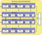
Automatic Age Estimation and Majority Age Classification from Multi-factorial MRI Data

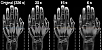
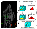
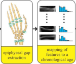
Age estimation from radiologic data is an important topic both in clinical medicine as well as in forensic applications, where it is used to assess unknown chronological age or to discriminate minors from adults. In this work, we propose an automatic multi-factorial age estimation method based on MRI data of hand, clavicle and teeth to extend the maximal age range from up to 19 years to up to 25 years. Fusing age-relevant information from all three anatomical sites, our method utilizes a deep convolutional neural network that is trained on a dataset of 322 subjects in the age range between 13 and 25 years, to achieve a mean absolute prediction error in regressing chronological age of 1.01+/-0.74 years.

Stern D, Payer C, Giuliani N, Urschler M: Published in IEEE Journal of Biomedical and Health Informatics, 23(4):1392-1403, 2019. PDFDOI (open access)

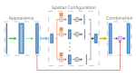
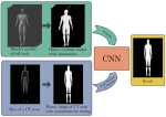
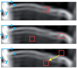
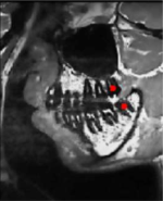
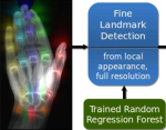
Integrating spatial configuration into heatmap regression based CNNs for landmark localization

In many medical image analysis applications, only a limited amount of training data is available due to the costs of image acquisition and the large manual annotation effort required from experts. Training recent state-of-the-art machine learning methods like convolutional neural networks (CNNs) from small datasets is a challenging task. In this work on anatomical landmark localization, we propose a CNN architecture that learns to split the localization task into two simpler sub-problems, reducing the overall need for large training datasets. Our fully convolutional SpatialConfiguration-Net (SCN) learns this simplification due to multiplying the heatmap predictions of its two components and by training the network in an end-to-end manner.

Payer C, Stern D, Bischof H, Urschler M: Published in Medical Image Analysis 54:207-219, 2019. DOI (open access)

Reducing acquisition time for MRI-based forensic age estimation Radiology-based estimation of a living person’s unknown age has recently attracted increasing attention due to large numbers of undocumented immigrants entering Europe. To avoid the application of X-ray-based imaging techniques, magnetic resonance imaging (MRI) has been suggested as an alternative imaging modality. Unfortunately, MRI requires prolonged acquisition times, which potentially represents an additional stressor for young refugees. To eliminate this shortcoming, we investigated the degree of reduction in acquisition time that still led to reliable age estimates. Neumayer B, Schloegl M, Payer C, Widek T, Tschauner S, Ehammer T, Stollberger R, Urschler M: Published in Scientific Reports 8:2063, 2018. DOI (open access)
Integrating geometric configuration and appearance information into a unified framework for anatomical landmark localization In approaches for automatic localization of multiple anatomical landmarks, disambiguation of locally sim- ilar structures as obtained by locally accurate candidate generation is often performed by solely includ- ing high level knowledge about geometric landmark configuration. In our novel localization approach, we propose to combine both image appearance information and geometric landmark configuration into a unified random forest framework integrated into an optimization procedure that iteratively refines joint landmark predictions by using the coordinate descent algorithm. Depending on how strong multiple land- marks are correlated in a specific localization task, this integration has the benefit that it remains flexible in deciding whether appearance information or the geometric configuration of multiple landmarks is the stronger cue for solving a localization problem both accurately and robustly. Urschler M, Ebner T, Stern D: Published in Medical Image Analysis 43:23-36, 2018. DOI (open access),BAA Web X-Ray Landmark annotations
Multi-Factorial Age Estimation from Skeletal and Dental MRI Volumes Age estimation from radiologic data is an important topic in forensic medicine to assess chronological age or to discriminate minors from adults, e.g. asylum seekers lacking valid identification documents. In this work we propose automatic multi-factorial age estimation methods based on MRI data to extend the maximal age range from 19 years, as commonly used for age assessment based on hand bones, up to 25 years, when combined with wisdom teeth and clavicles. Stern D, Kainz P, Payer C, Urschler M: Pupblished in Machine Learning in Medical Imaging. MLMI 2017. Lecture Notes in Computer Science, vol 10541. Springer, Cham. PDF, DOI
Forensic Age Estimation by Morphometric Analysis of the Manubrium from 3D MR Images Forensic age estimation research based on skeletal structures focuses on patterns of growth and development using different bones. In this work, our aim was to study growth-related evolution of the manubrium in living adolescents and young adults using magnetic resonance imaging (MRI), which is an image acquisition modality that does not involve ionizing radiation. Martinez-Vera N, Hoeller J, Widek T, Neumayer B, Ehammer T, Urschler M: Published in Forensic Science International 277:21-29, 2017. PDF,DOI
Regressing Heatmaps for Multiple Landmark Localization Using CNNs We explore the applicability of deep convolutional neural networks (CNNs) for multiple landmark localization in medical image data. Exploiting the idea of regressing heatmaps for individual landmark locations, we investigate several fully convolutional 2D and 3D CNN architectures by training them in an end-to-end manner. We further propose a novel SpatialConfiguration-Net architecture that effectively combines accurate local appearance responses with spatial landmark configurations that model anatomical variation. Payer C, Stern D, Bischof H, Urschler M: Presented at International Conference on Medical Image Computing and Computer Assisted Intervention (MICCAI) 2016, Athens, Greece. PDF, DOI
Automated Age Estimation from Hand MRI Volumes Using Deep Learning Biological age (BA) estimation from radiologic data is an important topic in clinical medicine, while in legal medicine it is employed to approximate chronological age. In this work, we propose the use of deep convolutional neural networks (DCNN) for automatic BA estimation from hand MRI volumes, inspired by the way radiologists visually perform age estimation using established staging schemes that follow physical maturation. Stern D, Payer C, Lepetit V, Urschler M: Presented at International Conference on Medical Image Computing and Computer Assisted Intervention (MICCAI) 2016, Athens, Greece. PDF, DOI
From Local to Global Random Regression Forests: Exploring Anatomical Landmark Localization State of the art anatomical landmark localization algorithms pair local Random Forest (RF) detection with disambiguation of locally similar structures by including high level knowledge about relative landmark locations. In this work we pursue the question, how much high-level knowledge is needed in addition to a single landmark localization RF to implicitly model the global configuration of multiple, potentially ambiguous landmarks. Stern D, Ebner T, Urschler M: Presented at International Conference on Medical Image Computing and Computer Assisted Intervention (MICCAI) 2016, Athens, Greece. PDF, DOI
Automatic localization of locally similar structures based on the scale-widening random regression forest Selection of set of training pixels and feature range show to be critical scale-related parameters with high impact on results in localization methods based on random regression forests (RRF). We present a scale-widening RRF method that effectively handles such ambiguities. Stern D, Ebner T, Urschler M: Presented at International Symposium on Biomedical Imaging (ISBI) 2016, Prague, CZ. PDF, DOI
From individual hand bone age estimates to fully automated age estimation via learning-based information fusion Increasingly important for both clinical and forensic medicine, radiological age estimation is performed by fusing independent bone age estimates from hand images. In this work, we show that the artificial separation into bone independent age estimates as used in established fusion techniques can be overcome. Thus, we treat aging as a global developmental process, by implicitly fusing developmental information from different bones in a dedicated regression algorithm. Stern D, Urschler M: Presented at International Symposium on Biomedical Imaging (ISBI) 2016, Prague, CZ. PDF, DOI
Anatomical landmark detection in medical applications driven by synthetic data An important initial step in many medical image analysis applications is the accurate detection of anatomical landmarks. Most successful methods for this task rely on data-driven machine learning algorithms. However, modern machine learning techniques, e.g. convolutional neural networks, need a large corpus of training data, which is often an unrealistic setting for medical datasets. In this work, we investigate how to adapt synthetic image datasets from other computer vision tasks to overcome the underrepresentation of the anatomical pose and shape variations in medical image datasets. Riegler G, Urschler M, Rüther M, Bischof H, Stern D: Presented at International Conference on Computer Vision Workshop TASKCV (ICCVW) 2015, Santiago de Chile, Chile. PDF, DOI
What automated age estimation of hand and wrist MRI data tells us about skeletal maturation in male adolescents Age estimation of individuals is important in human biology and has various medical and forensic applications. The aim of this paper was to investigate how informative automatically selected image features are regarding their ability to discriminate age, by exploring a recently proposed software-based age estimation method for MR images of the left hand and wrist. Urschler M, Grassegger S, Stern D: Published in Annals of Human Biology 42(4):356-65, 2015. PDF,DOI
Automatic third molar localization from 3D MRI using random regression forests Radiological age estimation of living subjects from MR images has recently become very popular. Besides skeletal ossification this can be done using the mineralization status of wisdom teeth. We propose a random regression forest framework to localize third molars in challenging 3D MRI datasets. Unterpirker W, Ebner T, Stern D, Urschler M: Presented at 19th Conference on Medical Image Understanding and Analysis (MIUA) 2015, Lincoln, UK. PDF
Fully Automatic Bone Age Estimation from Left Hand MR Images There has recently been an increased demand in bone age estimation (BAE) of living individuals and human remains in legal medicine applications. We propose a completely automated method for BAE based on volumetric hand MRI images. We see this work as a promising first step towards a novel MRI based bone age estimation system, with the key benefits of lacking exposure to ionizing radiation and higher accuracy due to exploitation of volumetric data. Stern D, Ebner T, Bischof H, Grassegger S, Ehammer T, Urschler M: Presented at International Conference on Medical Image Computing and Computer Assisted Intervention (MICCAI) 2014, Boston, US. PDF,DOI
Towards Automatic Bone Age Estimation from MRI: Localization of 3D Anatomical Landmarks Bone age estimation (BAE) is an important procedure in forensic practice which recently has seen a shift in attention from Xray to MRI based imaging. To automate BAE from MRI, localization of the joints between hand bones is a crucial first step, which is challenging due to anatomical variations, different poses and repeating structures within the hand. We propose a landmark localization algorithm using multiple random regression forests, first analyzing the shape of the hand from information of the whole image, thus implicitly modeling the global landmark configuration, followed by a refinement based on more local information to increase prediction accuracy. Ebner T, Stern D, Donner R, Bischof H, Urschler M: Presented at International Conference on Medical Image Computing and Computer Assisted Intervention (MICCAI) 2014, Boston, US. PDF,DOI
Determination of legal majority age from 3D magnetic resonance images of the radius bone The determination of an individual’s legal majority age is becoming increasingly important in forensic practice. We propose an automatic 3D method for the determination of legal maturity from MR images based on the ossification of the radius bone. Age estimation is performed by a linear regression model of the epiphyseal gap volume over the known ground truth age of training data. Stern D, Ebner T, Bischof H, Urschler M: Presented at International Symposium on Biomedical Imaging (ISBI) 2014, Beijing, China. PDF