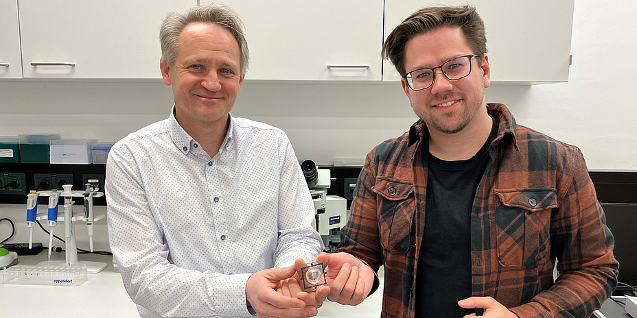
Researchers at the Institute of Health Care Engineering with European Testing Center of Medical Devices at Graz University of Technology (TU Graz) are devoting their work to the human heart, among other things. They use a special technology that allows non-invasive examination of cardiac activity in a petri dish using a microchip called microelectrode arrays.
Microelectrode arrays
Microelectrode arrays (MEAs) consist of several microelectrodes in close proximity to each other. They can be attached to the surface of a petri dish and connected to high-resolution measurement electronics. Thanks to different technologies, several hundred to several thousand electrodes can be fitted on just one square millimetre. These arrays can both stimulate the heart cells and take measurements.
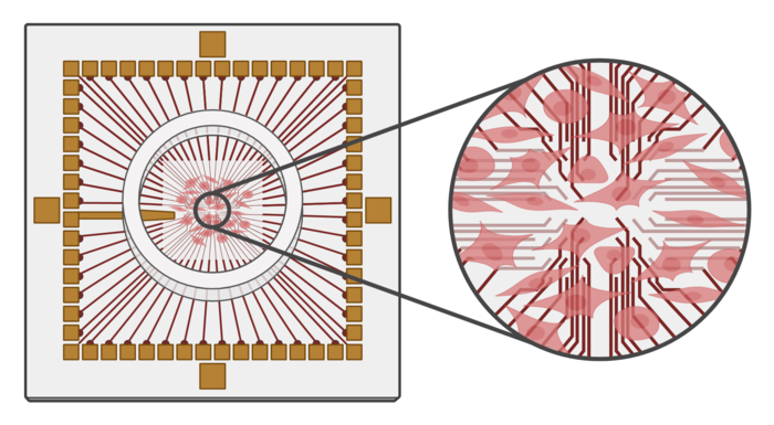
A microelectrode array with cultured heart muscle cells (created with BioRender.com).
Heart muscle cells are cultivated on these petri dishes to simulate cardiac activity. Within a few days, they combine to form a dense cell layer and special pacemaker cells that periodically send electrical signals to the cells in their vicinity. These signals, known as action potentials, change the cell membrane potential in several phases. In the very fast depolarisation phase, positive sodium ions flow into the cell, and the cell membrane potential increases. In the plateau phase, positively charged calcium ions flow out of the cell. And during the final repolarisation phase, positively charged potassium ions finally flow out of the cell and the potential returns to the original resting membrane potential. This signal is usually recorded using patch clamp measurements, in which pointed pipettes are inserted into cells to measure the potential changes.
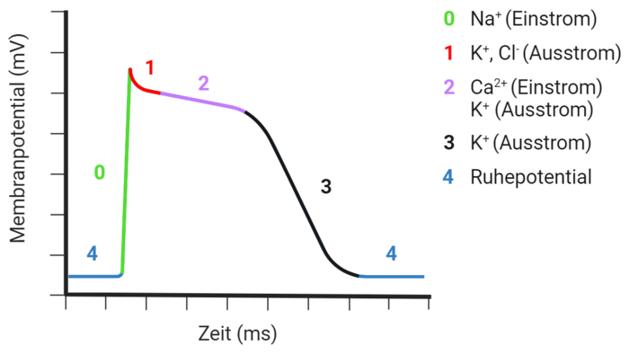
Phases of the action potential of a heart muscle cell (created with BioRender.com).
Measurements of the action potential using MEA electrodes
In contrast to this invasive method, MEA electrodes can also measure the signals in the form of an extracellular field potential on the surface of the cell. When a cardiac muscle cell triggers an action potential, it spreads across the cultivated cell layer in the petri dish and also activates the neighbouring cells; they contract in a way similar to a natural heartbeat. The electrode array measures this spread of activity across the measurement field. As the cells are not damaged during the measurement, the course of activity in the cell culture can be measured over several days or even weeks.
MEA technology is particularly suitable for investigations in the field of arrhythmia, for drug tests or research into the effects of new active ingredients.
Examinations of heart arrhythmia
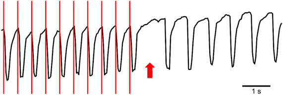
Heart damage, stress or ischaemia can disrupt the periodic heart rhythm and lead to arrhythmic contractions, which in turn can have serious and even life-threatening consequences. The heart itself has various mechanisms to stabilise the heart rhythm again. One of these is the so-called overdrive suppression, which is triggered when heart cells are stimulated with frequencies that are higher than their intrinsic beat frequency. Because the intracellular and extracellular ion concentrations cannot return to a normal level between stimuli due to the high frequency, there is a temporary pause in electrical activity. This stimulation can be simulated by MEAs and its consequences investigated. Different stimulation protocols influence the length of the activity pause, depending on the duration, intensity and number of stimuli.
These research activities are carried out jointly with the European Testing Center of Medical Devices at the Institute of Health Care Engineering at TU Graz (Daniel Ziesel, Sonja Langthaler, Theresa Rienmüller, Christian Baumgartner), the Department of Medical Physics and Biophysics (Brigitte Pelzmann, Klaus Zorn-Pauly) and the Clinical Department of Paediatric Cardiology at the Medical University of Graz (Daniela Baumgartner, Hannes Sallmon).
Christian BAUMGARTNER
Univ.-Prof. Dipl.-Ing. Dr.techn.
Institute of Health Care Engineering with European Testing Center of Medical Devices
Tel.: +43 316 873 7377
christian.baumgartner@tugraz.at