Images for download at the end of the message.
In most cases, aortic dissection is the result of a tear in the inner layer of the aortic wall, the intima. This causes blood to rush into the resulting space, forming what is known as a false lumen, which fundamentally changes the mechanical strain on the aortic wall. In the course of an aortic dissection, this can lead to aneurysmal expansion of the false lumen, which ruptures through the aortic wall with fatal consequences. A blood clot may also form in the false lumen, which in the best case fills the newly created passageway, thereby averting more severe effects. Although the condition can have serious implications, the reasons behind it have not yet been fully explained, and it is difficult to make an accurate prognosis.
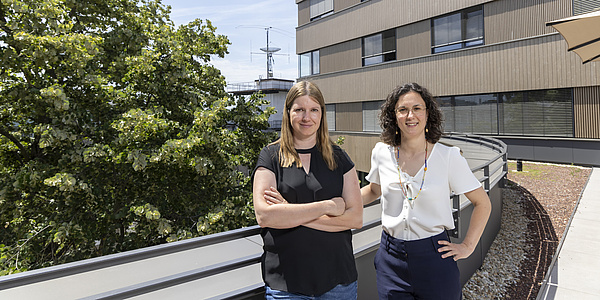
Over the past three years, researchers at TU Graz have been working to shed light on these questions as part of a cross-faculty lead project entitled Mechanics, Modeling and Simulation of Aortic Dissection. Their goal is to unravel the causes and progression of aortic dissection using IT-supported biomechanical methods. The project, which was recently extended for three years following a successful interim hearing, has now given rise to a separate research focus, and the plan is to continue work in this field after the extension expires.
TU Graz’s lead projects initiative provides funding for outstanding scientific, multidisciplinary research proposals. Funding is provided to projects selected by an independent, international jury. Once approved, a lead project is allocated around EUR 2m over three years, and this can be extended by a further three years following a successful evaluation, again performed by an independent, international jury. Two further lead projects are currently under way, entitled Dependable Internet of Things in Adverse Environments and Porous Materials@Work.
Successful initial phase
The lead project team has recorded a number of significant achievements in the past three years.
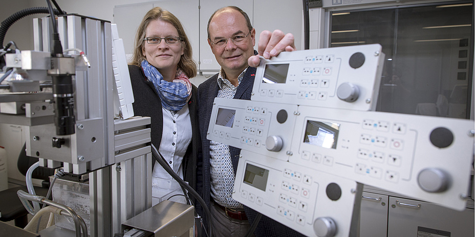
For example, the researchers have developed a new non-invasive method that uses available imaging methods; this has simplified the diagnosis and medical monitoring of aortic dissection. Simulations of blood flow dynamics have shown that this method allows for more precise prognosis of the condition and examination of the status of a dissection.
Detailed mechanical tests have also been carried out on human tissue – these have highlighted differences in the mechanical and structural properties of healthy and diseased tissue, such as the tensile strength, rigidity and the distribution of proteins in the aortic wall. Material models of the aortic wall were developed on the basis of these findings, which are designed to produce clearer insights into the causes and progression of aortic dissection with the help of sophisticated computer models. When performing the modelling experiments, the researchers also found explanations for the formation of blood clots in the false lumen and for the way in which blood flow dynamics influences the progression of aortic dissection. Ultimately, the lead project team developed software that enables them to reconstruct the initial state of the aorta prior to dissection, which in turn can be used for prognosis.
A copy of the press release on the project launch is available online.
Plans for second project phase
The recently approved second phase of the project will build on these encouraging results. The interdisciplinary team is aiming for a more detailed understanding of the causes and progression of aortic dissection, as well as developing artificial intelligence technologies based on Bayes’ theorem of probability that can predict the course of the condition. To do this, they intend to analyse existing clinical data (big data) in order to identify the underlying cause of acute aortic dissection or its relationship with other anomalies in the aorta. Subsequently, this will pave the way for predicting indications of the need for surgical restructuring as well as the progression of the condition, which in turn will allow for primary preventive, pre-clinical treatment.
The overriding goal for the coming three years is to bring more hospitals and companies on board, as well as establishing the new research centre and turning it into an integral component of biomedical research in Graz.
This research area is anchored in the Field of Expertise “Human & Biotechnology”, one of five strategic foci of TU Graz.
Gerhard A. HOLZAPFEL
Univ.-Prof. Dipl.-Ing. Dr.techn.
Institute of Biomechanics
Stremayrgasse 16/II
8010 Graz
Phone: +43 316 873 35500
holzapfel@tugraz.at
www.biomech.tugraz.at