Photoemission electron microscopy

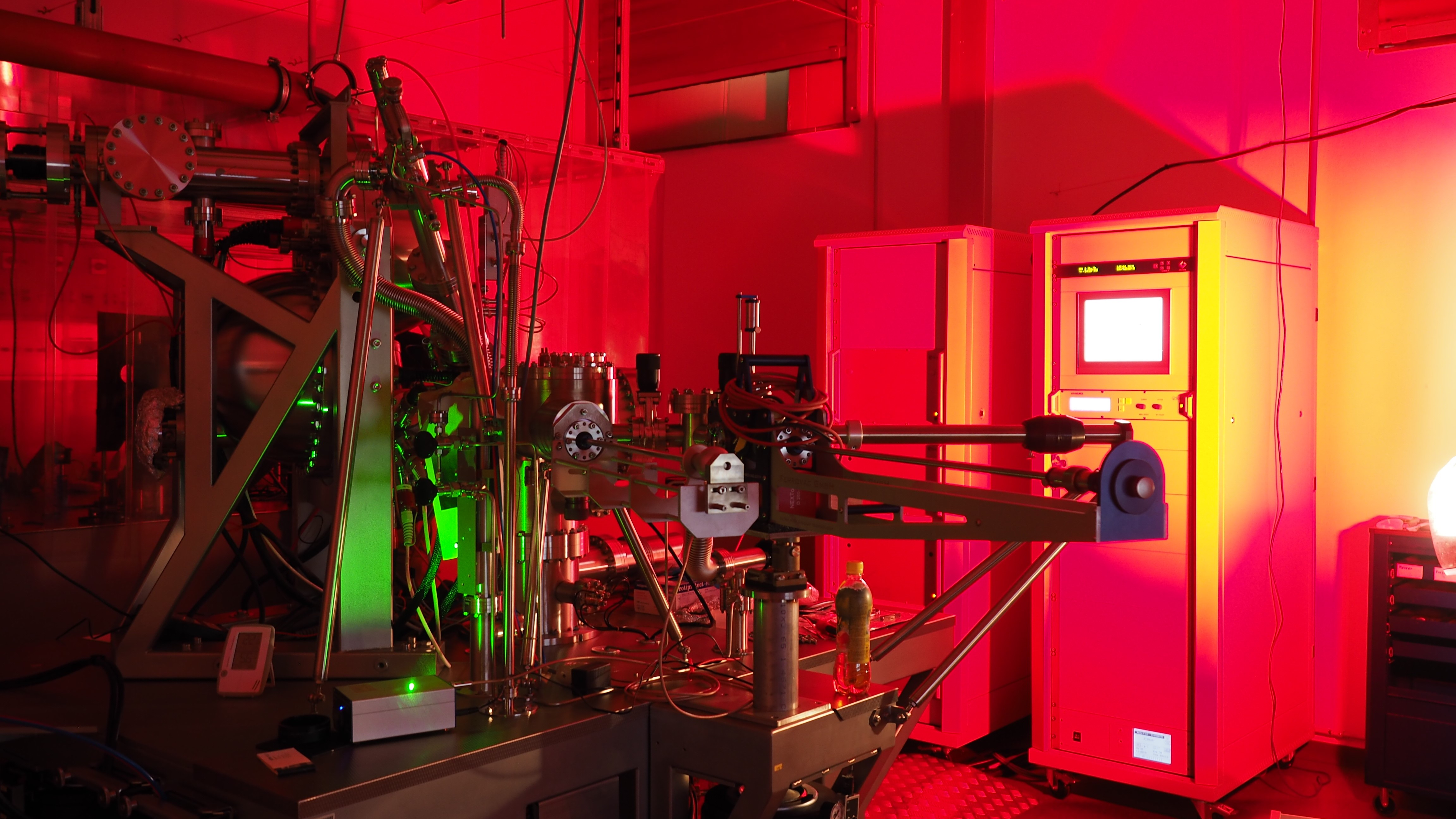
Photoemission electron microscopy (PEEM) visualizes the local variation of electrons emitted from a surface with high spatial resolution. We employ various light sources to generate photoelectrons ranging from Hg and He discharge lamps to nanosecond and femtosecond Ti:Sapphire lasers in order to explore the properties of materials at the nanoscale. The NanoESCA microscope in our laboratory is equiped with a special double hemispherical energy filter (EF), allowing for photoelectron spectroscopy with spatial resolution below 100 nm and k-space microscopy. Examples of our current research encompass the investigation of plasmonic nanostructures and nanoparticles as well as quantum materials. 


[-] d-bands of Cu(110) recorded with the NanoESCA in k-space microscopy mode.

Momentum space microscopy with our NanoESCA instrument allows us to map the band structure of materials. An example can be seen in the animated image, showing k-space images as a function of the kinetic energy of the electrons (in this case: EF = 21.3 eV) recorded with the NanoESCA and the He discharge lamp as light source. 

All images © TU Graz/Institute of Experimental Physics
Group members
image/svg+xml


Prof. Martin SCHULTZE
schultzenoSpam@tugraz.at
+43 (316) 873-8142


Em. Univ.-Prof. Wolfgang E. ERNST
wolfgang.ernstnoSpam@tugraz.at
+43 (316) 873-8140

 
Thomas JAUK
thomas.jauknoSpam@student.tugraz.at
+43 (316) 873-8144