Photoemission electron microscopy

Photoemission electron microscopy (PEEM) visualizes the local variation of electrons emitted from a surface with high spatial resolution. We employ various light sources to generate photoelectrons ranging from Hg and He discharge lamps to nanosecond and femtosecond Ti:Sapphire lasers in order to explore the properties of materials at the nanoscale. The NanoESCA microscope in our laboratory is equiped with a special double hemispherical energy filter (EF), allowing for photoelectron spectroscopy with spatial resolution below 100 nm and k-space microscopy. Examples of our current research encompass the investigation of plasmonic nanostructures and nanoparticles as well as quantum materials. 


d-bands of Cu(110) recorded with the NanoESCA in k-space microscopy mode. Click on the image to start the animation.

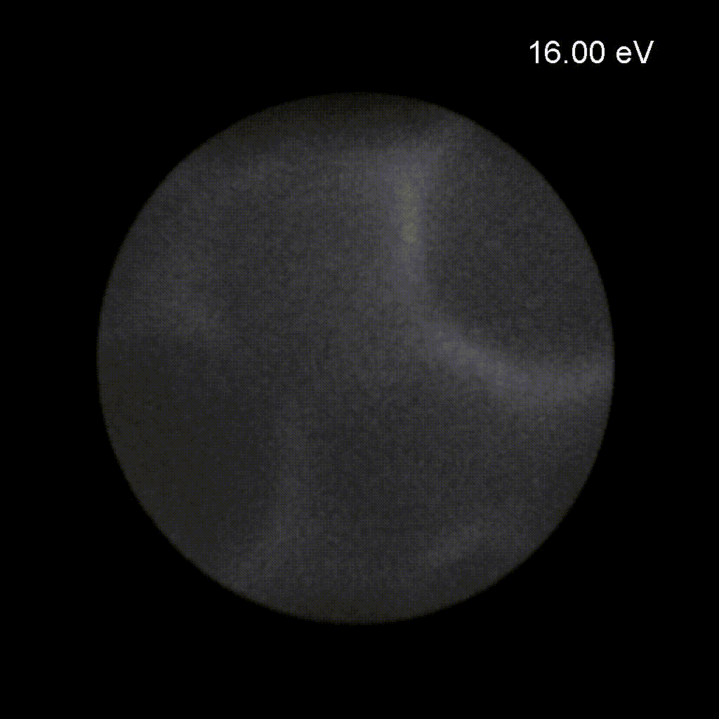
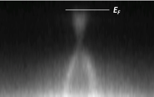
Momentum space microscopy with our NanoESCA instrument allows us to map the band structure of materials. An example can be seen in the animated image, showing k-space images as a function of the kinetic energy of the electrons (in this case: EF = 21.3 eV) recorded with the NanoESCA and the He discharge lamp as light source. 

All images © TU Graz/Institute of Experimental Physics
Group members
image/svg+xml

 
Dr. Florian LACKNER
florian.lacknernoSpam@tugraz.at
+43 (316) 873-8666


Prof. Martin SCHULTZE
schultzenoSpam@tugraz.at
+43 (316) 873-8142
orcid-ID


Prof. Wolfgang E. ERNST
wolfgang.ernstnoSpam@tugraz.at
+43 (316) 873-8140


DI Alexander SCHIFFMANN
alexander.schiffmannnoSpam@tugraz.at
+43 (316) 873-8667

 
Thomas JAUK
thomas.jauknoSpam@student.tugraz.at
+43 (316) 873-8144