There is a plethora of applications for electrical stimulation of excitable cells, be it for research, therapy or diagnostic purposes. Various research projects and established technologies aim to stimulate cells artificially in order to elicit a response or improve cellular growth, proliferation and interaction. This can lead to direct responses at a cellular level, as well as functional improvements over time.
The work for this thesis will mainly focus on the electrical stimulation of neurons and networks of neurons, including optoelectronic stimulation with the help of a novel technology called photocapacitor. When a photocap is stimulated by an external light source, the resulting potential change at its surface causes the neuronal cell membrane to depolarize, possibly leading to an action potential. The aim is to find and test suitable stimulation protocols to change the plasticity of connected neurons and the rate of spontaneous activity, which could eventually be used to mitigate or even reverse damages to brain tissue caused by traumatic brain injury (TBI).
To establish a theoretical basis, the efficacy of different stimulation methods and protocols that are already used in experimental practice need to be researched. Their suitability for TBI rehabilitation and their relevance for direct electrical stimulation with electrodes or photocapacitors will be evaluated. Several techniques are then selected to conduct further experiments with in vitro samples and mathematical models.
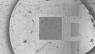
Experiments will initially be done with primary cultures of dissociated neurons from neonatal rat hippocampus and cortex. The major advantages of these samples are that they are relatively easy to maintain, can survive for long periods of time in an incubator and the setup required to keep them alive during measurements is relatively simple. Later on, organotypic hippocampal slice cultures will be used to examine the effect of established stimulation protocols on tissue that shows characteristics similar to in vivo brain tissue. Finally, hippocampal slices from rat brains that were experimentally subjected to TBI will be stimulated and the resulting changes to neuronal plasticity and spontaneous activity analyzed. The regions of the hippocampal structure and the interactions of the neurons therein are very well known, making it an ideal candidate to investigate detrimental factors caused by TBI.
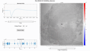
Signals will be measured with the help of multielectrode arrays (MEAs) systems and patch clamping setups to determine neuronal activity on a cellular level as well as the synchronized activity within a network of neurons. These systems can also be used to apply electrical stimuli of various shapes to the samples and acquire the direct response and changes over longer periods of time. Optoelectronic stimulation may be induced by MEAs with small photocaps applied between the electrode pads or by culturing the neurons on a transparent conductive material (e.g. ITO) with a larger photocap layer.
A model of the electrical activity within networks of neurons and their reaction to stimuli shall be developed to make in silico testing of stimulation protocols possible. Structure and parameters of this model will be determined with the help of existing research and cell models, as well as insights gathered through experiments. A model of the neuronal interactions within the hippocampal structure may serve as a starting point for simulations of a hippocampus damaged by TBI. The results from stimulation protocols applied to these models should ultimately be compared with the experimental data gathered from measurements with neuronal samples.