MR Image Reconstruction
Our group develops new methods for Magnetic Resonance Imaging (MRI) with the goal of faster, more robust, and quantitative imaging. Our research interests extend from the development of fundamentally new measurement techniques to the translation of new methods into clinical use. Methodologically, we mostly focus on computational imaging methods that combine advanced numerical algorithms for image reconstruction with jointly designed data acquisition techniques.
Maschine Learning Image Reconstruction
We investigate the use of generative AI for use as prior knowledge in a Bayesian formulation of image reconstruction (Luo et al. 2023). Recently, we started to work on self-supervised machine learning techniques that can learn to improve MRI scans even when there is no ground truth avaiable (Blumenthal et al. 2024).
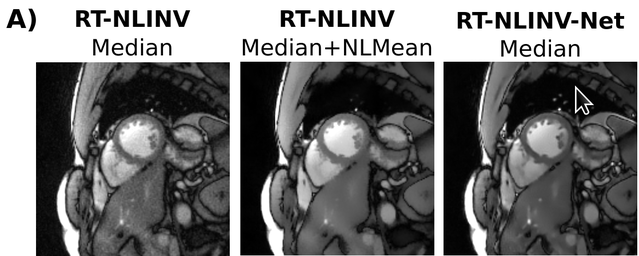
Figure: A comparison of different reconstruction techniques for real-time MRI. RT-NLINV-Net is our newest maschine learning technique that can learn without ground truth images (Blumenthal et al. 2024).
References:
- Moritz Blumenthal, Chiara Fantinato, Christina Unterberg-Buchwald, Markus Haltmeier, Xiaoqing Wang, Martin Uecker. Self Supervised Learning for Improved Calibrationless Radial MRI with NLINV-Net. Magnetic Reosnance in Medicine, early view (2024)
- Guanxiong Luo, Moritz Blumenthal, Martin Heide, Martin Uecker. Bayesian MRI Reconstruction with Joint Uncertainty Estimation using Diffusion Models. Magnetic Resonance in Medicine 90:295-311 (2023)
- Moritz Blumenthal, Guanxiong Luo, Martin Schilling, H. Christian M. Holme, Martin Uecker. Deep, Deep Learning with BART. Magnetic Resonance in Medicine 89:678-693 (2023)
Calibrationless Parallel MRI with ENLIVE
ENLIVE is our new algorithm for efficient and robust calibrationless parallel MRI. ENLIVE is based on NLINV but also integrates an important feature inspired by ESPIRiT: The classical SENSE model is relaxed to make the algorithm more robust to model violations.
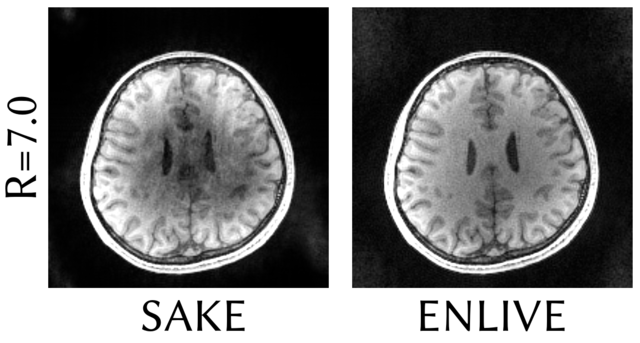
Figure: A comparison of ENLIVE vs SAKE as another calibrationless parallel imaging method which is based on structured low-rank matrix completion. Reconstruction time when using a single core was 22 seconds for ENLIVE and 6.3 hours using SAKE (Holme et al. 2019).
References:
- H. Christian M. Holme, Sebastian Rosenzweig, Frank Ong, Robin N. Wilke, Michael Lustig, Martin Uecker. ENLIVE: An Efficient Nonlinear Method for Calibrationless and Robust Parallel Imaging. Scientific Reports 9:3034 (2019) arXiv:1706.09780 [physics.med-ph]
Autocalibrated Parallel MRI with ESPIRiT
ESPIRiT is an algorithm for autocalibrated parallel MRI, which combines the robustness of the GRAPPA method with the speed and flexibility of a SENSE-based reconstruction methods. The corresponding publication with Peng Lai (GE Healthcare), Michael Lustig (UC Berkeley) and colleagues is the highest-cited research paper published in the year 2014 (after two years) in Magnetic Resonance in Medicine, the leading jounral in MR methodology. Implementations of ESPIRiT calibration and reconstruction are available in our reconstruction toolbox.
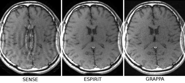
Figure: Images of a human brain acquired with a small FOV. While the SENSE reconstruction has an artifact in the center, GRAPPA is free from this problem. Using multiple set of maps estimated with the ESPIRiT calibration method, a SENSE-based ESPIRiT reconstruction is able to produce an artifact-free image similar to GRAPPA (Uecker et al. 2014).
References:
- Martin Uecker, Michael Lustig. Estimating Absolute-Phase Maps Using ESPIRiT and Virtual Conjugate Coils Magnetic Resonance in Medicine 77:1201-1207 (2017) PMC5018407 arXiv:1509.03557 [cs.CV]
- Martin Uecker, Peng Lai, Mark J. Murphy, Patrick Virtue, Michael Elad, John M. Pauly, Shreyas S. Vasanawala, Michael Lustig. ESPIRiT - An Eigenvalue Approach to Autocalibrating Parallel MRI: Where SENSE meets GRAPPA. Magnetic Resonance in Medicine 71:990-1001 (2014) PMC4142121 [source code: Reconstruction Toolbox] <mark>highest cited paper published in Magnetic Resonance in Medicine in 2014</mark>
Calibrationless Parallel MRI with Nonlinear Inverse Reconstruction
Hiqh quality reconstruction in parallel MRI requires exact knowledge of the sensitivity profiles of the receive coils. In nonlinear inverse reconstruction, image content and coil sensitivities are estimated jointly, which avoids an explicit calibration step and improves reconstruction quality especially if the amount of calibration data is small. The problem leads to a blind-deconvolution problem (although the roles of frequency and time are switched in MRI). Because the technique can be applied directly to non-Cartesian data, it is ideal for real-time MRI with radial data acquisition. In fact, our method for real-time MRI is based on this algorithm.
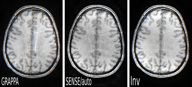
Figure: 3D FLASH MRI with 2D acceleration of 4 = 2x2 and 16x16 reference lines. Comparison between GRAPPA, SENSE with coil sensitivities obtained from the fully sampled k-space center, and nonlinear inverse (Inv) reconstruction (Uecker et al. 2008).
References:
- Sebastian Rosenzweig, H. Christian M. Holme, Robin N. Wilke, Dirk Voit, Jens Frahm, Martin Uecker. Simultaneous Multi-Slice Reconstruction Using Regularized Nonlinear Inversion: SMS-NLINV. Magnetic Resonance in Medicine, 79:2057-2066 (2018) arXiv:1705.04135 [physics.med-ph] <mark>Gö-VIP-16 Clinical Science Award</mark>
- Florian Knoll, Christian Clason, Kristian Bredies, Martin Uecker, Rudolf Stollberger. Parallel Imaging with Nonlinear Reconstruction using Variational Penalties. Magnetic Resonance in Medicine 67:34-41 (2012) [free full text] PMC4011127 [PubMan] [source code] <mark>2011 Stefan-Schuy-Award, Austrian Society for Biomedical Engineering, Winner</mark>
- Martin Uecker, Shuo Zhang, Jens Frahm. Nonlinear Inverse Reconstruction for Real-time MRI of the Human Heart Using Undersampled Radial FLASH. Magnetic Resonance in Medicine 63:1456-1462 (2010) [source code]
- Martin Uecker, Thorsten Hohage, Kai Tobias Block, Jens Frahm. Image Reconstruction by Regularized Nonlinear Inversion - Joint Estimation of Coil Sensitivities and Image Content. Magnetic Resonance in Medicine 60:674-682 (2008) [free full text] [PubMan] [source code] <mark>Gorter Award, Finalist</mark>
(Non-Cartesian) Parallel MRI with Compressed Sensing
Compressed sensing is a new technique that can be used to accelerate measurements by exploiting the redundancy (compressibility) of the acquired data. Our publication from 2007 is one of the first examples where this method is used in an imaging application. For more information about compressed sensing in MRI, see this page from Michael Lustig at UC Berkeley who pioneered the use of this technique in MRI. Parallel MRI and compressed sensing can be combined to achieve even higher acceleration for MRI, which is the basis for most advanced image reconstruction methods in MRI. The 2007 paper is the first work to use this important combination.
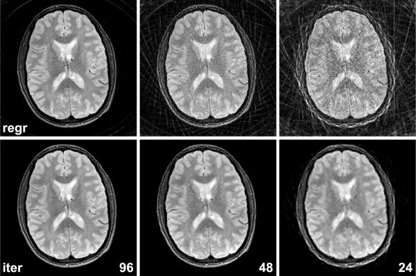
Figure: Reconstruction of a human brain from 96, 48, and 24 radial spokes. (Top) Conventional gridding reconstruction (Bottom) Combination of linear non-Cartesian parallel imaging (generalized SENSE) and compressed sensing using a total-variation penalty (Block et al. 2007).
References:
- Florian Knoll, Christian Clason, Kristian Bredies, Martin Uecker, Rudolf Stollberger. Parallel Imaging with Nonlinear Reconstruction using Variational Penalties. Magnetic Resonance in Medicine 67:34-41 (2012) [free full text] PMC4011127 [PubMan] [Matlab code] <mark>2011 Stefan-Schuy-Award, Austrian Society for Biomedical Engineering, Winner</mark>
- Kai Tobias Block, Martin Uecker, Jens Frahm. Undersampled Radial MRI with Multiple Coils. Iterative Image Reconstruction Using a Total Variation Constraint Magnetic Resonance in Medicine 57:1086-1098 (2007) [free full text] [PubMan] <mark>2007 ISMRM Young Investigator (I.I. Rabi) Award, Winner</mark>
Total Generalized Variation

Fig.1: Radial Brain Scan: Clinical 3T Scanner, 32 Receiver Coils, 256x256 Matrix, 32 (top row) and 24 (bottom row) radial projections. Left: Conventional reconstruction method with streaking artifacts. Right: Image reconstruction with a Total Generalized Variation (TGV) constraint (Knoll et al. 2011)
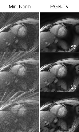
Fig.2: Radial Cardiac Real-Time-FLASH Scan: Clinical 3T Scanner, 32 Receiver Coils, 128x128 Matrix, 55 (top row), 28 (middle row) and 19 (bottom row) radial projections, corresponding to temporal resolutions of 110ms, 56ms and 38ms.
Left: Conventional reconstruction method with streaking artifacts.
Right: Nonlinear Parallel Imaging Reconstruction with a variational constraint (IRGN-TV, Knoll et al. 2012)
References:
- Florian Knoll, Christian Clason, Kristian Bredies, Martin Uecker, Rudolf Stollberger. Parallel Imaging With Nonlinear Reconstruction Using Variational Penalties. Magnetic Resonance in Medicine 67:34 - 41 (2012)
- Florian Knoll, Kristian Bredies, Thomas Pock, Rudolf Stollberger. Second Order Total Generalized Variation (TGV) for MRI. Magnetic Resonance in Medicine 65:480-491 (2011)
- Florian Knoll, Christian Clason, Clemens Diwoky, Rudolf Stollberger. Adapted Random Sampling Patterns for Highly Accelerated MRI. Magnetic resonance materials in physics, biology and medicine 24:43 - 50 (2011)
- Florian Knoll, Gerrit Schultz, Kristian Bredies, Daniel Gallichan, Maxim Zaitsev, Jürgen Hennig, Rudolf Stollberger. Reconstruction of undersampled radial PatLoc imaging using total generalized variation. Magnetic Resonane in Medicine 70:40-52 (2013)
- Florian Knoll, Unger, M.; Clemens Diwoky, Christian Clason, Thomas Pock, Rudolf Stollberger. Fast reduction of undersampling artifacts in radial MR angiography with 3D total variation on graphics hardware. Magnetic resonance materials in physics, biology and medicine 23:103 - 114 (2010)