
Confocal laser scanning microscopy (CLSM)
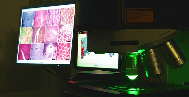
We are equipped with a Leica TCS SPE laser scanning confocal microscope, able to sequentially analyze up to 8 channels for fluorescent light plus one transmitted-light channel. The microscope includes also a Leica DFC360FX camera for recording non-confocal, filter based fluorescent light.
Images in Gallery
Growth chamber for plants
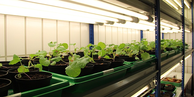
We establish model systems for studying plant-associated bacteria and fungi, their beneficial effects on the plant growth and their efficiency in the biocontrol of plant pathogens.
Gaschromatograph-Mass Spectrometer
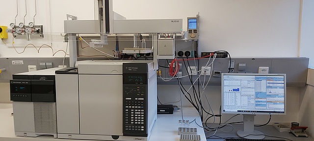
With the GC-MS System from the company Agilent, equipped with the PAL-System the analysis of liquid, volatile and gaseous samples is possible.
For the detection of microbial volatile organic compounds (mVOCs) we use the method of Solid Phase Microextraction (SPME).
Liquid Chromatography with High Accurate Mass Analyzer
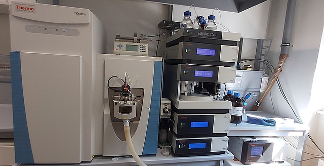
As a core member of the Central Lab, located at the Karl Franzens University, we use the UHPLC-Orbitrap MS Analyzer mainly for our metabolomic approach. The data analysis is done with the software "Compound Discoverer" of Thermofisher.