


Quantitative CT-derived vessel metrics in idiopathic pulmonary fibrosis: A structure function study
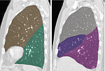
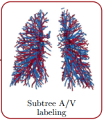
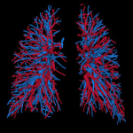
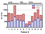
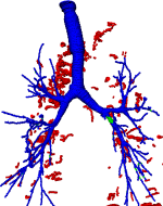
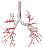
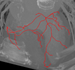
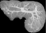
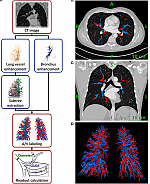
Knowledge of the lung vessel morphology in healthy subjects is necessary to improve our understanding about the functional network of the lung and to recognize pathologic deviations beyond the normal inter-subject variation. In order to determine morphologic readouts from a large number of healthy subjects, computed tomography pulmonary angiography (CTPA) datasets, negative for pulmonary embolism, and other thoracic pathologies, were analyzed using a fully-automatic, in-house developed artery/vein separation algorithm. Our fully-automatic artery/vein separation algorithm provided reliable measures of pulmonary arteries and veins with respect to age and gender. There was a large variation between subjects in all readouts. No relevant dependence on age, gender, or vessel type was observed. These data may provide reference values for morphometric analysis of lung vessels.
Jacob J, Pienn M, Payer C, Urschler M, Kokosi M, Devaraj A, Wells A W, Olschewski H: Published in Respirology 24(5): 445-452, 2019. DOI